Published by GM Optical | Expert Vision Care Insights
What Is Retinitis Pigmentosa?
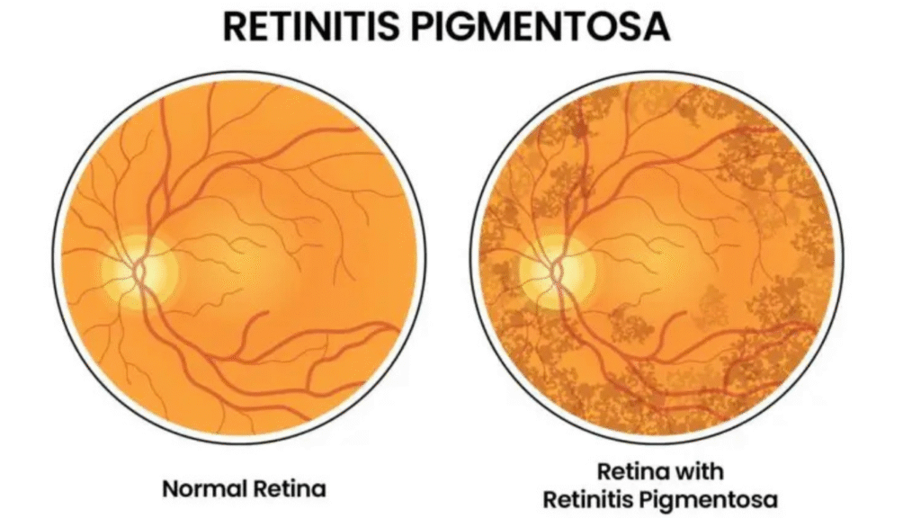
Retinitis Pigmentosa (RP) describes a cluster of rare, hereditary conditions that impact the retina—the light sensitive layer at the back of the eye that plays a crucial role in the process of seeing. RP conditions ultimately cause a slow degeneration of the retina and consequent vision loss. RP is rare, affecting only about 1 in 4,000 individuals globally, but it is still one of the most common causes of genetic vision loss.
Causes and Risk Factors
Retinitis Pigmentosa is genetic in origin. Mutations in more than 60 different genes can lead to the condition, each affecting how retinal cells function or maintain themselves.
It can be inherited in several ways:
- Autosomal dominant RP – passed from one affected parent, typically a milder form.
- Autosomal recessive RP – occurs when both parents carry the gene, though they may not show symptoms.
- X-linked RP – primarily affects males, as the mutation lies on the X chromosome.
Early Signs and Symptoms
RP usually begins in childhood or adolescence, though the rate of progression varies between individuals. Common symptoms include:
- Night blindness – difficulty seeing in dim lighting or darkness.
- Tunnel vision – gradual loss of peripheral (side) vision.
- Difficulty adapting to light changes – sensitivity to bright lights or glare.
- Loss of color perception – colors may appear faded over time.
- Central vision loss – occurs in later stages, affecting reading and facial recognition.
Because symptoms progress slowly, regular eye examinations are crucial for early detection and monitoring.
Diagnosis
Diagnosis is typically made through specialized eye tests, such as:
- Visual field testing – to measure peripheral vision.
- Electroretinography (ERG) – to evaluate the electrical activity of retinal cells.
- Optical Coherence Tomography (OCT) – to visualize retinal structure in detail.
- Genetic testing – to identify specific gene mutations and assess family risk.